
What is arthroscopic discopexy?
Discopexy means repositioning and securing the TMJ disc. You may also hear this called meniscopexy or meniscus repair; here, “meniscus” refers to the TMJ disc. Through small openings beside the ear, a camera and fine instruments allow the surgeon to work inside the joint. The disc is mobilized, moved into a more useful position, and held with sutures or another fixation method chosen for the joint.
This follows principles used in orthopedic arthroscopy: work through small openings, preserve the joint’s cushioning tissue when possible, and repair or stabilize it rather than remove it. The TMJ disc serves a cushioning role similar to a knee meniscus, although the anatomy and repair are different.
A joint washout removes fluid and inflammatory material. Discopexy adds disc repositioning and fixation. The aim is to improve joint mechanics, pain, and movement; it does not guarantee a normal disc position or a pain-free joint.
Symptoms, examination, and usually MRI, CBCT, or both guide the decision. Surgery addresses a problem inside the joint. Muscle pain may still need conservative treatment.
During surgery
- Anesthesia and access. Your anesthesia plan is tailored to the procedure and your health and discussed before surgery. Small access points are made beside the ear, with a small incision just inside the ear to help secure the sutures. One or both joints may be treated.
- Inspect and wash the joint. The camera shows the disc, joint surfaces, inflammation, and scar tissue. The joint is irrigated with sterile fluid.
- Free and reposition the disc. A controlled cut releases the attachment of the upper part of the lateral pterygoid muscle at the front of the disc when needed. This reduces forward pull and helps the disc move back. Scar tissue that limits movement may also be released. The disc is then repositioned and secured.
- Finish the repair. Joint movement is checked and the small access sites are closed. Dr. Yim usually injects regenerative material into the joint during the procedure. The material used varies with the joint findings and treatment plan. Its purpose, potential benefits, and limitations are discussed before surgery.
If the disc is badly torn or the joint is too damaged, the planned repair may not be possible. The alternatives and possibility of further surgery should be discussed beforehand.
Surgical photos and videos
Real operative images from Dr. Yim’s teaching presentations. The close-up views show surgical tissue and may include blood. Videos are silent and do not play automatically.
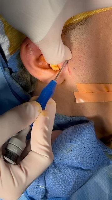
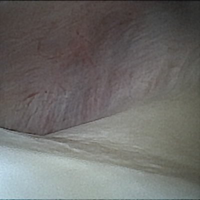
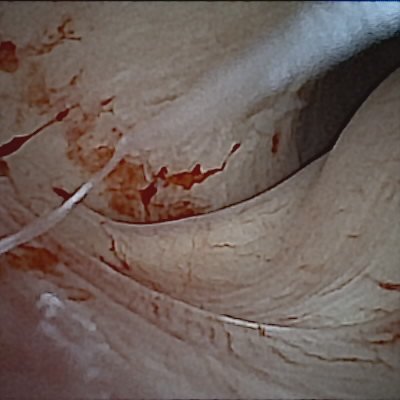
Introducing the arthroscope
Treatment inside the joint
Risks
- Pain, swelling, bruising, bleeding, infection, stiffness, and small scars.
- Facial nerve injury can cause weakness of facial movement, including difficulty closing an eye. This is usually temporary; rarely, weakness or paralysis can be lasting. Other nerve injuries may cause numbness.
- Ear-canal or eardrum injury, ear fullness, dizziness, or hearing changes. Serious damage to the structures of the middle ear is extremely uncommon but possible and can cause lasting hearing loss. Hearing may stay the same or worsen. Tinnitus is not expected to reliably improve; hearing concerns warrant an ENT assessment and hearing test.
- A changed bite, which may require an appliance, orthodontics, or occasionally further surgery.
- Persistent pain or clicking, recurrent disc displacement, failure of fixation, or a need for another procedure. Muscle-related pain can remain even when the joint repair succeeds.
- Anesthetic complications. Extremely rare but serious complications include injury through the thin bone separating the joint from the skull cavity, with possible bleeding, leakage of fluid around the brain, or brain injury. Injury to nearby blood vessels and fluid swelling around the airway are also possible.
Your own risk depends on the joint, the procedure, and your health. This guide supplements the discussion and consent process at your visit.
Recovery and time off
The first few days
Many patients go home the same day. Arrange a ride and an adult to stay with you after anesthesia. Expect some swelling, soreness, and stiffness. Follow your discharge instructions for medication, wound care, and cold packs.
Work and school
Plan for 3–4 days off work or school. Some patients need up to one week. Your job, symptoms, and the extent of surgery may affect when you can return. Returning to class or work does not mean the disc has finished healing.
Do not drive, operate machinery, or make important decisions during the restriction period given by your anesthesia team, or while taking sedating pain medicine.
Eating, exercise, and jaw movement
Choose soft foods for comfort while eating is sore. Avoid opening your mouth too wide for the first six weeks. Do not force a stretch or start opening exercises on your own.
Jaw exercises usually begin at 6–8 weeks, after follow-up with Dr. Yim. He will check your healing and tell you which movements to start. Movement and comfort can improve over weeks to months. Keep follow-up appointments to check the bite, opening, pain, and healing; imaging may be repeated when useful.
Appliances and orthodontics
An appliance may be used during recovery. Dr. Yim commonly recommends an orthodontic evaluation for arthroscopic surgery patients to coordinate the bite after joint treatment. The timing is individualized; braces are not an automatic requirement for every patient.
When to get help
Seek emergency care for trouble breathing or swallowing, rapidly increasing swelling, or uncontrolled bleeding. Contact your surgical team promptly for fever, worsening pain or swelling, drainage, a new hearing change, or difficulty closing an eye.
Dr. Yim’s usual recovery schedule is listed above. Sources for general surgical information: Royal Free London: TMJ arthroscopy · BAOMS: TMJ arthroscopy · Sah et al.: arthroscopic discopexy outcomes · Lateral pterygoid release study · Middle-ear complications · Rare neurological complications. Published arthroscopy schedules may differ from your disc-fixation instructions. Follow the written plan from your surgical team.